PCV / haematocrit
The proportion of blood made up of red cells. It is checked against the dog’s own history and the laboratory reference interval. A value is never read in isolation from hydration, clinical examination and the rest of the CBC.
Screening protects two animals: the dog who gives and the patient who receives. This page explains what we examine, what each laboratory group contributes, why PCR matters, and how preventive care is kept current.
Start with the whole dog
Enrolment begins with history, records and a physical assessment. We review previous illness, medication, transfusion history, travel, tick exposure, diet, preventive care, temperament and the family’s ability to return.
Before every collection we repeat the history and examine temperature, weight trend, hydration, mucous membranes, pulse, respiration, heart, lungs, lymph nodes, skin, coat and venepuncture sites. A dog can pass every previous test and still be deferred on the day.
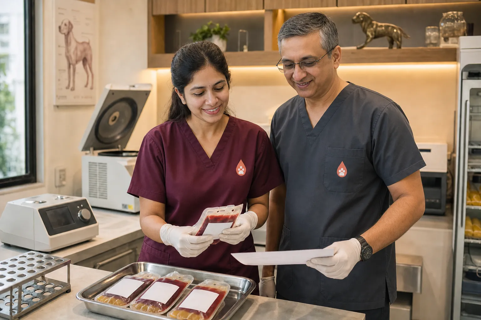
Complete blood count
A CBC is not just a haemoglobin result. We review cell numbers, indices, morphology and the relationships between them.
The proportion of blood made up of red cells. It is checked against the dog’s own history and the laboratory reference interval. A value is never read in isolation from hydration, clinical examination and the rest of the CBC.
Two further measures of circulating red-cell mass and oxygen-carrying capacity. Agreement between PCV, haemoglobin and the red-cell count helps identify inconsistent or artefactual results.
MCV describes average red-cell size; MCHC describes average haemoglobin concentration; RDW describes variation in cell size. Together they help characterise anaemia and flag a result that needs investigation before donation.
Young red cells released by the marrow. The absolute reticulocyte count helps show whether the marrow is responding appropriately when red-cell mass is low.
Total leukocytes plus neutrophils, lymphocytes, monocytes, eosinophils and basophils. The pattern can point to inflammation, infection, stress, allergy or another process that needs clarification.
Platelets are central to primary haemostasis. We review the numerical count, platelet size indices and the blood film because platelet clumping can make an automated count look falsely low.
A microscope check of red-cell shape and colour, white-cell morphology, platelet estimate, clumps and visible blood parasites. The film is the quality check on the machine-generated numbers.
LFT, KFT and biochemistry
Panels are groups of related measurements. No single enzyme proves normal organ function, and an out-of-range result is interpreted with the dog, sample quality, medication, hydration and previous values.
| Group | Measurements | What we learn |
|---|---|---|
| Liver-associated enzymes | ALT, ALP and, where included, AST and GGT | These are interpreted as patterns, not as a single pass-or-fail 'liver test'. ALT can rise with hepatocellular injury; ALP can rise with cholestasis, steroid influence, bone activity and other conditions. An abnormality is investigated rather than labelled from one number. |
| Liver function and protein balance | Bilirubin, albumin, total protein, globulin, glucose and cholesterol | These help assess excretion, protein synthesis and wider metabolic context. Albumin and total protein also matter when interpreting plasma quality and the donor’s hydration. |
| Renal filtration markers | Urea / BUN, creatinine and SDMA where available | Urea is influenced by hydration, diet, bleeding and liver function. Creatinine is influenced by renal filtration and muscle mass. SDMA can add information where creatinine alone may be insensitive. Results are read together. |
| Electrolytes and minerals | Sodium, potassium, chloride, calcium and phosphorus where included | These help identify dehydration, losses, endocrine or renal abnormalities and sample problems that need resolving before a dog donates. |
| Urinalysis | Specific gravity, dipstick chemistry, sediment and protein assessment | Urine concentration and sediment add information that blood urea and creatinine cannot provide alone. Protein or active sediment may prompt repeat sampling or further renal investigation. |
Tick-borne disease PCR
PCR is used because a healthy-looking dog can carry a transmissible organism and a routine smear can miss low-level infection. The exact panel is reviewed against Mumbai epidemiology, travel and exposure.
Molecular screen
Canine babesiosis can cause haemolysis and may be present before obvious illness. Molecular testing looks for pathogen DNA; species-level reporting, when available, helps interpretation.
Molecular screen
A brown-dog-tick-associated infection that may be subclinical and can affect platelets, marrow and other systems. PCR complements the examination, CBC and exposure history.
Molecular screen
The panel is selected for locally relevant organisms, including platelet-associated infection where applicable. A platelet count forms part of the same clinical picture.
Molecular screen
A tick-associated protozoal infection relevant in many warm regions. PCR may be combined with careful blood-film review and exposure history.
PCR amplifies a selected section of pathogen genetic material from the submitted sample. A positive result therefore means the target was detected; it is not inferred from antibody exposure and it is not based only on seeing an organism on a smear.
The organism must be present in the sampled material above the assay’s detection limit. Low circulating burden, intermittent circulation, prior antimicrobial or antiprotozoal treatment, collection timing, transport and inhibition can all affect detection.
The selected target was not detected in that sample at that time. It does not prove that the dog has never been exposed, does not cover organisms outside the panel, and does not replace history, examination, CBC, film review or repeat testing after a new exposure.
Organism load, timing, prior treatment, sample handling and assay design affect molecular detection. Results are interpreted with the exposure history, examination, CBC, platelet count and blood film. Testing is repeated according to risk and before a dog returns after a relevant gap or exposure.
Blood type and other checks
DEA 1 typing is recorded so recipient matching begins with known donor information. Coagulation testing, faecal testing, urinalysis and additional infectious-disease assays are added when history, geography or examination makes them relevant.
Prevention programme
Every donor leaves enrolment with a written preventive-care plan. The schedule follows the dog, the product and local risk—not a generic calendar copied onto every record.
At enrolment we review the complete vaccine record, not just the date of the last injection. Core canine protection covers distemper virus, adenovirus and parvovirus. Rabies vaccination follows applicable law and the licensed product. Leptospirosis and other non-core vaccines are selected for local exposure and lifestyle. The veterinarian records what is due, what is not due, and why.
For dogs with a documented puppy series, core doses should have continued at two- to four-week intervals until at least 16 weeks of age, followed by a booster at or after 26 weeks. Thereafter, modified-live core vaccines are generally given no more frequently than every three years. The annual clinical review still happens, and the exact schedule follows the licensed product and the dog’s record.
Rabies vaccination follows Indian law, local requirements and the licensed product; dates and certificate details stay in the donor record. Where leptospirosis vaccination is indicated, a two-dose primary course is required and revaccination is generally annual because protection is shorter-lived than modified-live core vaccination.
The plan considers age, diet, scavenging, outdoor access, household animals and previous parasite findings. A suitable broad-spectrum product is given at its labelled interval, and adult donors undergo periodic faecal testing rather than relying on tablets alone. A positive result is treated and rechecked before donation.
Protection is continuous because brown dog ticks can persist in homes and kennels. Depending on the dog, the veterinarian may select an oral isoxazoline such as a fluralaner-, afoxolaner-, sarolaner- or lotilaner-type product, or a suitable topical spot-on or collar. Each has a different labelled interval; the written schedule follows the exact product used.
A preventive cannot compensate for an infested environment. Families are shown how to check the dog, wash bedding, address all household pets and seek professional environmental treatment when brown dog ticks have established indoors.
A dog can have missed doses, prior exposure or a subclinical infection. Preventive treatment reduces risk; history, examination, CBC, film review and PCR are still required for donor safety.
“Spot-on”, “tablet” and “collar” are delivery types, not schedules. Fluralaner, afoxolaner, sarolaner, lotilaner and topical products do not all share the same interval or contraindications. We record the active ingredient, brand, dose, date given, next due date and any adverse effect. A missed or uncertain dose is treated as a gap, not rounded up as current.
When we defer
Fever, weight loss, dehydration, medication, recent illness, vaccination timing, parasite exposure, an unexpected laboratory result, anxiety or a change in temperament can all pause donation. Deferral is not failure and it is never overridden because a unit is wanted.
Send us your dog’s age, breed, weight, medical history and current preventive-care record. We will explain the assessment before anything is booked.